The eye is an amazing optical device. Like a camera, the eye focuses light into a clear image, which is then changed into electrical signals and sent to the brain for interpretation. Abnormalities in the size and shape of the eye lead to poor vision, which requires glasses or contact lenses for improved clarity.
Scientists and engineers have been working for years, trying to perfect the options for restoring vision without glasses or contact lenses. Currently, the two major categories of vision enhancement include LASIK/PRK and ICL lens implant procedures. LASIK and PRK work externally by reshaping the cornea, ICL implants are placed inside the eye to help focus light rays without glasses or contact lenses. Each procedure has specific benefits and minor risks. Our job is to help educate you on your options and guide your decision making so that you end up with the best possible vision with the safest and highest technology procedure.
Dr. DeBry will work with each patient to make sure they get the best technology for their vision-correction surgery.
A generally healthy eye and cornea
No uncontrolled dry eye symptoms
Adequate corneal thickness – measured at your pre-op with a safe and simple test (pachymetry)
Eyelid and facial structures that allow the laser to get near the eye
Dissatisfaction with glasses and/or contact lenses
A desire to have improved vision with a willingness to accept some risk to achieve this goal
Success in vision-correction surgery requires high-quality standards and attention to fine details. Dr. Peter W. DeBry brings these qualities to Las Vegas with his extensive training in eye surgery and lens implantation. Dr. DeBry completed medical school at the University of Utah in 1996. At that time he was honored with induction into the prestigious Alpha Omega Alpha medical honor society, reserved for the top physicians in each graduating class. Next, he spent three years of residency focused on medical and surgical eye care in Madison, Wisconsin, where he was chosen to help coordinate resident training as the chief resident. After this, he was one of only four doctors nationally selected to attend one of the country’s top fellowships in eye surgery techniques and spent the next year at the Bascom Palmer Eye Institute in Miami, Florida. After working with top doctors learning the most up-to-date surgical techniques, he moved to Kansas City, Missouri where he taught these techniques to new eye surgeons as Associate Clinic Professor at the University of Missouri. Finally, he relocated in 2003 to help provide cataract, and vision correction surgery for the residents of Las Vegas.
Dr. DeBry is one of the most experienced eye surgeons in Las Vegas. From performing a corneal transplant in a 90-year-old to a vision-saving glaucoma surgery in a baby, he does more complex surgeries than any other eye surgeon. Dr. DeBry was invited to be among the first few groups of eye surgeons in the United States trained in the surgical techniques for Verisyse and Staar Visian ICL Lens implantation. He has been doing these procedures since 2005, longer than any other full-time surgeon in Las Vegas. He has presented research at local and national meetings and has published articles in national ophthalmology journals.
At NV Eye Surgery, our LASIK surgeons use the most advanced LASIK technology available to deliver truly customized vision correction treatments tailored to your unique eyes. We use two advanced laser systems that work together to reshape your corneas with personalized precision.
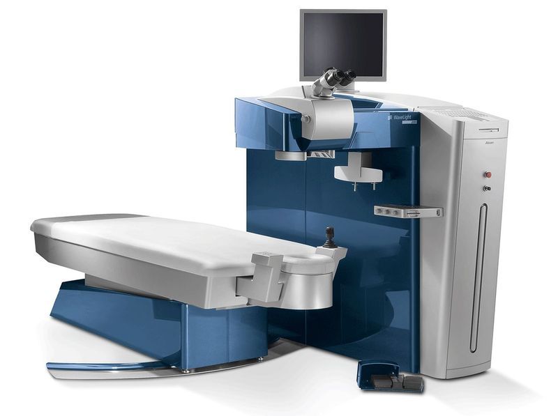
The Alcon Wavelight EX500 Laser sculpts the corneas through a fast, computer-controlled laser, which is accurate to a tiny fraction of a millimeter. Its rapid pulses ensure maximum safety and effectiveness as it makes precise changes to the surface of your cornea.
Our LASIK surgeons also use the Ziemer LDV Z4 Femto Laser, which creates a detailed topographic map of your cornea. This helps guide the laser in customizing subtle adjustments according to your eyes’ natural contours. Using both of these advanced lasers that provide mapping and precision enables an ultra-customized reshaping for spectacular vision outcomes tailored to you.
Our Contoura Vision platform takes LASIK personalization further than ever before. Unlike standard LASIK, which relies on simple wavefront mapping technology to guide treatments, Contoura is customized specifically and precisely to your unique eye topography.
Using advanced mapping, this technology can capture thousands of data points across your corneas to fully understand every subtle curve that makes your eyes one-of-a-kind. Your LASIK surgeon can then use this detailed blueprint of your eyes to guide the laser in reshaping your corneas in a way that smooths out imperfections and improves your vision tailored specifically to you.
LASIK at NV Eye Surgery in Las Vegas, Nevada, with Contour Vision, is customized and fine-tuned to the natural structure of your eyes only. The benefit is the best possible visual outcomes with incredible sharpness, vivid color, and ultra-clear night vision.
Contoura Vision is a type of LASIK surgery that creates a detailed map of your cornea, the clear outer layer of your eye. During LASIK with Contoura Vision, your LASIK surgeon uses the Contoura Vision system to create a detailed map of your eye and then inputs that information into the laser to reshape the cornea according to your unique map.
This precisely smooths out any uneven areas on the cornea, removing any irregularities and improving your eyes’ ability to focus. Contoura Vision works exceptionally well if you have both nearsightedness and astigmatism, which is when the cornea has an irregular curve. The highly personalized Contoura Vision system leads to excellent visual results.
As described previously, LASIK treatments require a thin flap to be made on the cornea. In the past, this flap was created with a very sharp blade called a microkeratome. In a small percentage of people, the microkeratome-created unique problems as the flap were cut with imperfections or centering issues. To lower the risk of irregular flaps a laser was developed for flap creation. This Femtosecond laser cuts tissue with an accuracy of 0.01 mm and makes perfectly circular flaps with a very consistent thickness. The use of all-laser LASIK has made LASIK a safer procedure. It has also added a little more cost to the procedure because now 2 lasers are used instead of one. An interesting side note, the femtosecond laser fires a laser pulse 0.000000000000001 seconds in duration. This rapid burst of energy causes a microscopic explosion in the tissue, breaking bonds.
Normal LASIK uses the refraction numbers based on your glasses to create a treatment plan. Studies have shown that vision can be improved and some negative visual symptoms decreased if the laser is programmed to treat higher order aberrations along with the refraction numbers. To achieve this, additional measurements are made on the eyes to measure the individual “fingerprint” of your eye. These measurements are then put into the LASIK excimer computer to provide a custom treatment designed just for you. This may give better results depending on your corneal structure and refraction.
Having eye surgery is a bit like a trip to the dentist. It is a little uncomfortable, takes about 15 minutes to have it done, and has great benefits. It can be scary as some people don’t like things coming up close to their eyes. To help relax you we will provide a prescription for relaxing medication, a sleeping pill commonly used to relax anxious nerves. If you are small in stature and not an anxious person taking ½ of a pill is usually fine. If you have a lot of anxiety or are a bigger person you can take a whole pill or in some cases even more.
During the procedure, you will lie down on a comfortable bed with a firm headrest. Get comfortable, as it is very important to hold still once the procedure starts. The eyelids are cleansed and sticky tape is put around the eyelashes to keep them out. A small wire is used to gently hold open the eyelids. During the treatment, there are several steps…
The Femtosecond laser will cut the flap. A suction ring is placed on the eye during this step. It takes only 15 seconds but is a bit uncomfortable due to the pressure from the suction ring. Your vision will fade to black during this time.
Next, the flap will be lifted and the corneal tissue prepared for the excimer laser. During this step, your vision will be blurry.
The excimer laser treatment is next. This takes between 15 and 45 seconds depending on the degree of correction being treated. There will be a red or green light during this time and it is important that you focus on the colored light to keep the laser treatment centered on the cornea.
After the laser, the flap will be irrigated. You will feel some cold water around the eye. Then the flap will be placed back into position and allowed to seal for a few minutes. The wire holding the lids will be removed and the treatment repeated on the other eye.
After the procedure, we encourage you to go home and take a nap. You will be sleepy from the relaxing medication and it is helpful to keep your eyes closed for a few hours to start the healing process. The vision will be a little blurry for the first 24 hours but will improve even more over the next several days as the cornea starts to heal.